A professional skin image analysis system contributes to clinical evaluation by replacing subjective human observation with quantitative, pixel-level data. By utilizing standardized lighting, high-resolution imaging, and specialized algorithms, these systems provide an objective measurement of both biological skin changes—such as melanin reduction—and the physical performance of the transdermal delivery device itself.
Core Takeaway The true value of professional image analysis lies in objectivity and sensitivity. It transforms qualitative clinical observations into hard data, allowing researchers to detect subtle therapeutic improvements and precise mechanical failures that traditional visual scoring would miss.
Quantifying Biological Efficacy
Eliminating Subjective Bias
In traditional dermatology studies, results often rely on a clinician's visual grading scale.
This method is inherently prone to variance between different observers.
Professional analysis systems remove this error by applying standardized light sources to ensure every image is captured under identical conditions, regardless of the operator.
Measuring Melanin Indices
To evaluate if a transdermal treatment is effectively delivering its active ingredients, researchers must look beyond surface redness.
Specialized software analyzes melanin indices and specific pigmented areas.
This allows for the precise tracking of hyperpigmentation reduction, quantifying exactly how much pigment has cleared over time.
Detecting Micro-Improvements
Human eyes struggle to perceive gradual, low-contrast changes in skin tone.
Digital algorithms can detect subtle improvements in skin quality that may not be immediately visible to the naked eye.
This sensitivity is critical for proving efficacy in early-stage trials or for treatments with cumulative, long-term effects.
Evaluating the Delivery System
Assessing Adhesion Accuracy
The efficacy of a transdermal treatment relies heavily on the patch staying in contact with the skin.
Image analysis software evaluates patch edges at a pixel level to calculate the exact percentage of remaining adhesion area.
This provides a far more accurate metric than manual visual estimates of lifting or peeling.
Visualizing Mechanical Failure
Physical stressors can cause the adhesive to shift, a phenomenon known as "cold flow."
Analysis systems can identify the "dark ring" formed by this adhesive flow.
By isolating these artifacts, researchers can distinguish between a dirty patch and actual mechanical failure.
Correlating Lab and Clinical Data
Laboratory mechanical tests do not always predict how a patch will perform on a moving human subject.
By quantifying wear performance digitally, researchers can build a stronger correlation between benchtop data and real-world clinical outcomes.
This helps engineers refine the physical design of the transdermal patch to ensure consistent drug delivery.
Understanding the Trade-offs
Complexity of Setup
Moving from visual scoring to digital analysis increases the complexity of the trial protocol.
Strict adherence to imaging standards is required; slight deviations in lighting or angle can skew pixel-level data.
Data Interpretation Requirements
While the system provides objective numbers, it yields a significantly higher volume of data than simple scoring sheets.
Teams must possess the expertise to interpret quantitative metrics correctly rather than relying on intuitive "before and after" comparisons.
Making the Right Choice for Your Goal
To maximize the value of a skin image analysis system, align your analysis method with your specific clinical endpoint.
- If your primary focus is Pharmacological Efficacy: Prioritize the measurement of melanin indices and pigmented areas to prove the active ingredient is biologically active.
- If your primary focus is Device Engineering: Concentrate on the quantification of adhesion area and edge analysis to validate the mechanical stability of the patch.
Objective data does not just describe the result; it validates the science behind the cure.
Summary Table:
| Evaluation Metric | Traditional Method | Image Analysis System Benefit |
|---|---|---|
| Biological Efficacy | Subjective visual grading | Quantitative melanin & pigment indices |
| Adhesion Stability | Manual visual estimates | Pixel-level calculation of contact area |
| Mechanical Failure | Qualitative observation | Precise detection of adhesive "cold flow" |
| Data Consistency | High observer variance | Standardized lighting and algorithmic precision |
Elevate Your Product Performance with Enokon's R&D Excellence
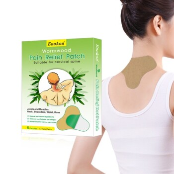
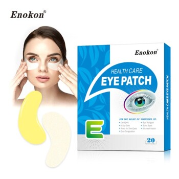
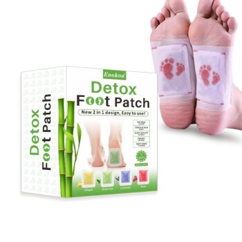
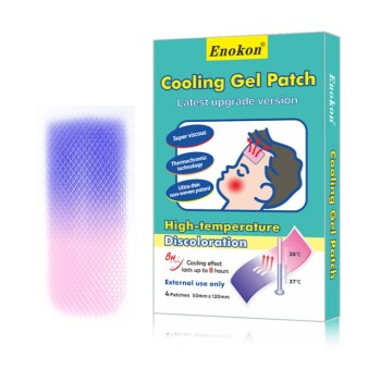
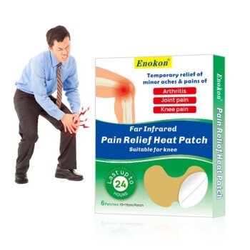
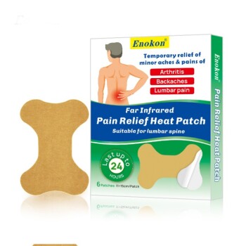
As a trusted manufacturer specializing in wholesale transdermal patches and custom R&D solutions, Enokon understands that clinical efficacy is the foundation of market success. We produce a comprehensive range of drug delivery products—including Lidocaine, Menthol, Capsicum, Herbal, and Far Infrared pain relief patches, as well as Eye Protection, Detox, and Medical Cooling Gel patches.
Our expertise goes beyond manufacturing; we provide the precision and quality control necessary to ensure your products meet the highest clinical standards (note: our capabilities exclude microneedle technology).
Ready to develop high-performance transdermal solutions? Contact us today to discuss how our custom R&D and manufacturing capabilities can bring your vision to life.
References
- Suyong Kim, Hyungil Jung. Enhanced Transdermal Delivery by Combined Application of Dissolving Microneedle Patch on Serum-Treated Skin. DOI: 10.1021/acs.molpharmaceut.7b00111
This article is also based on technical information from Enokon Knowledge Base .
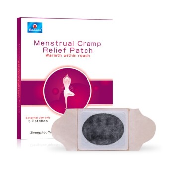
Related Products
- Silicone Scar Sheets Patch Transdermal Drug Patch
- Mugwort Wormwood Pain Relief Patch for Neck Pain
- Hydra Gel Health Care Eye Patch
- Far Infrared Heat Pain Relief Patches Transdermal Patches
- Icy Hot Menthol Medicine Pain Relief Patch
People Also Ask
- What are common adverse effects of transdermal drug delivery? Risks & Prevention Tips
- What is the role of methanol as a solvent in the extraction of active ingredients from Piper betle for transdermal patches?
- What role does a silicone-based transdermal delivery system play in Parkinson's? Enhancing Early-Stage Patient Care
- Why must modifications to preparation methods for transdermal drug delivery systems be documented in detail? - Essential Tips
- Where can users find more information about patch safety? Essential Guide to Safe Transdermal Use