Transmission Electron Microscopy (TEM) serves as the definitive visual validation tool for the morphological study of Huperzine A ethosomes. It provides high-resolution imaging that allows researchers to directly observe the physical form of the nanocarriers, confirming that they possess the necessary closed vesicular structure and near-spherical shape required for effective drug delivery.
Core Takeaway While particle size analyzers provide statistical data about the dimensions of a formulation, TEM offers the indispensable visual proof of its physical reality. It confirms that Huperzine A ethosomes have successfully formed into intact, closed systems capable of encapsulating the drug, rather than existing as random aggregates.
Visualizing the Nanostructure
Confirming Vesicular Shape
The primary contribution of TEM is the ability to resolve the microscopic morphology of the ethosomes.
By using high-energy electron beams, TEM allows you to clearly identify if the particles have achieved a near-spherical shape. This visual confirmation is critical because the shape of the nanocarrier directly influences how it interacts with biological membranes and delivers the Huperzine A payload.
Verifying Structural Integrity
Beyond simple shape, TEM allows for the inspection of the vesicle's architecture.
You are looking for a typical closed vesicular structure. This confirms that the lipid bilayer has successfully formed a continuous barrier, creating an enclosed internal space necessary for drug encapsulation. Without this closed structure, the formulation is merely a mixture of ingredients, not a functional delivery system.
Validating Quantitative Data
Grounding Indirect Measurements
Particle size analyzers and dynamic light scattering provide data on the average size of particles, but they cannot tell you what those particles look like.
TEM validates this indirect data. If your analyzer reports a specific particle size, TEM provides the visual evidence that these readings correspond to actual, intact vesicles rather than dust, broken membrane fragments, or amorphous clumps.
Detecting Aggregation
TEM provides a direct assessment of the formulation's dispersion quality.
It allows you to verify the absence of particle aggregation. Seeing individual, distinct vesicles confirms that the preparation process was successful and that the ethosomes are stable enough to remain separate, which is vital for consistent dosage and performance.
The Trade-off: Surface vs. Internal Structure
Understanding the Imaging limit
While TEM is superior for internal and through-structure imaging, it is important to understand its specific role compared to other methods.
TEM focuses on the internal microstructure and the arrangement of the phospholipid bilayer. However, it is often complementary to Scanning Electron Microscopy (SEM), which is better suited for observing macro-morphology and surface characteristics. Relying on TEM alone gives you deep structural insight but may miss broader surface topography; relying on SEM alone misses the critical bilayer confirmation.
Making the Right Choice for Your Goal
To effectively utilize TEM in your Huperzine A ethosome research, consider your specific objective:
- If your primary focus is Formulation Success: Use TEM to visually confirm the formation of closed, spherical bilayers, ensuring the lipids have self-assembled correctly.
- If your primary focus is Quality Control: Use TEM to detect aggregation and validate that the particle size distribution data matches the actual physical reality of the sample.
TEM is the bridge that transforms theoretical formulation data into proven structural reality.
Summary Table:
| Feature | Contribution of TEM in Ethosome Study | Impact on Research |
|---|---|---|
| Morphology | Confirms near-spherical shape | Validates delivery mechanism potential |
| Architecture | Verifies closed vesicular structure | Ensures successful drug encapsulation |
| Data Validation | Grounds indirect particle size data | Provides physical proof of statistical readings |
| Stability | Detects particle aggregation | Confirms formulation dispersion quality |
| Internal Detail | Resolves lipid bilayer arrangement | Proves structural integrity of the nanocarrier |
Elevate Your Product Innovation with Enokon
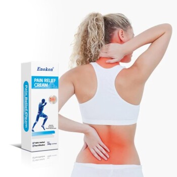
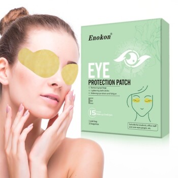
At Enokon, we understand that superior formulation is the foundation of effective delivery. As a trusted manufacturer specializing in wholesale transdermal patches and custom R&D solutions, we provide high-quality products ranging from Lidocaine, Menthol, and Capsicum pain relief to specialized Eye Protection, Detox, and Medical Cooling Gel patches.
While our expertise focuses on advanced transdermal drug delivery systems (excluding microneedle technology), we share your commitment to structural integrity and quality excellence. Whether you need reliable wholesale supply or a partner for custom product development, our team is ready to help you bring effective solutions to your customers.
Ready to scale your production? Contact us today to discuss how our manufacturing expertise can support your brand's growth.
References
- WU Ji-yu, Aifang Huang. Preparation and evaluation of transdermal permeation of Huperzine A ethosomes gel in vitro. DOI: 10.1186/s40360-024-00742-w
This article is also based on technical information from Enokon Knowledge Base .
Related Products
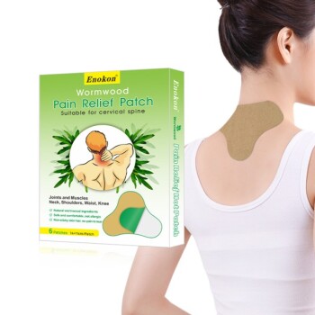
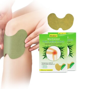
- Mugwort Wormwood Pain Relief Patch for Neck Pain
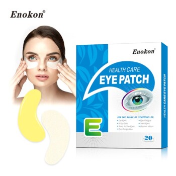
- Hydra Gel Health Care Eye Patch
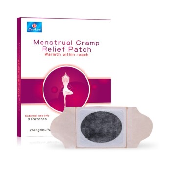
- Heating Pain Relief Patches for Menstrual Cramps
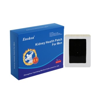
- Prostate Pain Kidney Health Care Patch for Men
- Natural Herbal Tube Cream for Pain Relief Analgesic Cream
People Also Ask
- What safety considerations should be kept in mind when using pain relief patches? Ensure Safe & Effective Pain Management
- Are pain relief pills and patches available without a prescription? Choose the Right OTC Pain Relief
- Who should consult a healthcare professional before using pain relief patches? Ensure Your Safety with Medical Advice
- How do pain relief patches compare to oral painkillers? Targeted Relief vs. Systemic Effects
- What type of pain are pain relief patches best suited for? Find Targeted Relief for Your Aches