High-resolution Scanning Electron Microscopy (SEM) serves as a critical diagnostic tool for identifying the physical causes of failure in transdermal formulations. It enables the direct visual monitoring of rate-controlling membranes, revealing failure points such as surface particle accumulation, pore degradation, and unwanted drug crystallization that quantitative data alone cannot detect.
By comparing membrane microstructure before and after permeation, SEM establishes a causal link between physical changes and performance failure. It reveals how specific morphological issues, such as smoothed pore edges or surface deposits, increase permeation resistance and compromise the drug delivery system.
Diagnosing Membrane Integrity and Function
Monitoring Microstructural Evolution
The primary application of SEM in this context is tracking the physical changes of the rate-controlling membrane throughout its lifecycle.
By imaging the membrane before and after permeation experiments, researchers can identify exactly when and how the structure degrades.
Detecting Surface Contamination and Blockage
Failures in drug delivery are often caused by physical barriers developing on the membrane surface.
SEM allows for the precise identification of surface deposits, specifically particle accumulation. These deposits often result from interactions with buffer components or the application of electrical current during iontophoresis.
Assessing Pore Morphology
The geometry of membrane pores is a decisive factor in drug release rates.
High-resolution imaging reveals subtle morphological changes, such as the smoothing of pore edges. These observations are vital because physical alterations to the pore structure directly increase the permeation resistance of the membrane.
Analyzing Drug Distribution and Stability
Identifying Crystallization vs. Dispersion
A common failure mode in transdermal patches is the physical instability of the drug within the polymer matrix.
SEM provides the resolution necessary to see if drug molecules have remained dispersed or if they have precipitated into crystals. Crystallization generally indicates a failure in formulation stability and effectively halts proper drug release.
Visualizing Internal Structures
Surface imaging alone is insufficient for a complete failure analysis.
SEM enables the observation of internal pore structures, providing insight into the tortuosity and connectivity of the matrix. This helps researchers understand the underlying mechanism of drug release—or the lack thereof.
Understanding the Trade-offs
Visual Data vs. Chemical Composition
While SEM is exceptional for identifying where a failure occurred (e.g., a deposit on a pore), it is a morphological tool, not a chemical one.
Visualizing a deposit confirms it is blocking a pore, but SEM alone may not identify the chemical composition of that deposit without auxiliary spectroscopy.
Surface Interpretation vs. Bulk Properties
SEM excels at surface and cross-sectional analysis, but it requires careful interpretation to ensure local defects represent the entire system.
Relying solely on a small visual sample area without correlating it to bulk permeation data can lead to incorrect conclusions about the formulation's overall failure mode.
Optimizing Formulations Based on Visual Data
To effectively use SEM for failure analysis, you must correlate visual defects with your specific performance goals.
- If your primary focus is Solving Low Permeation Rates: Investigate the membrane surface for particle accumulation or smoothed pore edges that are increasing resistance.
- If your primary focus is Improving Physical Stability: Examine the polymer matrix to ensure the drug remains amorphous and dispersed rather than crystallizing over time.
- If your primary focus is Membrane Selection: Use "before and after" imaging to discard membrane materials that show significant morphological degradation during testing.
Ultimately, SEM transforms failure analysis from a guessing game into a precise, evidence-based optimization process.
Summary Table:
| Failure Mode | SEM Application & Observation | Impact on Formulation |
|---|---|---|
| Pore Degradation | High-res imaging of pore edges and geometry | Increased permeation resistance |
| Surface Blockage | Detection of particle deposits/buffer residues | Reduction in drug delivery surface area |
| Crystallization | Visualization of drug precipitates vs. dispersion | Loss of stability and halted drug release |
| Structural Decay | "Before and after" microstructure comparison | Compromised membrane integrity |
| Internal Tortuosity | Cross-sectional analysis of pore connectivity | Unpredictable drug release mechanisms |
Optimize Your Transdermal Performance with Enokon
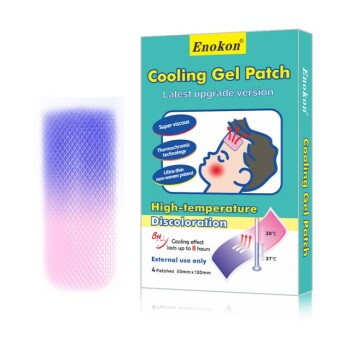
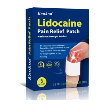
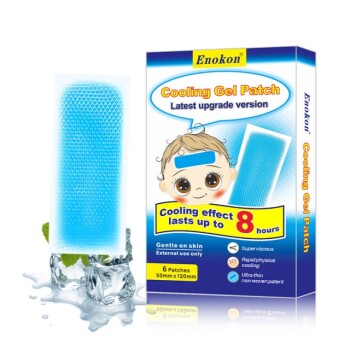
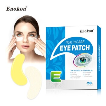
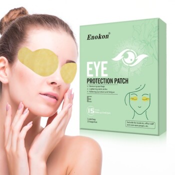
Don't let formulation failure slow down your R&D. Enokon is a trusted manufacturer and wholesale partner specializing in custom transdermal solutions. From Lidocaine, Menthol, and Capsicum pain relief to specialized Eye Protection and Medical Cooling Gel patches, we provide the R&D expertise and manufacturing precision needed to ensure your products meet the highest standards of stability and efficacy (excluding microneedle technology).
Ready to elevate your product line? Contact us today for custom R&D and wholesale solutions.
References
- Jia‐You Fang, Yi-Hung Tsai. Electrically-Assisted Skin Permeation of Two Synthetic Capsaicin Derivatives, Sodium Nonivamide Acetate and Sodium Nonivamide Propionate, via Rate-Controlling Polyethylene Membranes. DOI: 10.1248/bpb.28.1695
This article is also based on technical information from Enokon Knowledge Base .
Related Products
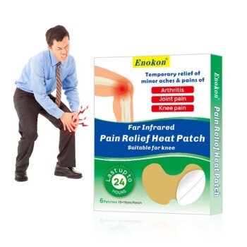
- Far Infrared Heat Pain Relief Patches Transdermal Patches
- Icy Hot Menthol Medicine Pain Relief Patch
- Silicone Scar Sheets Patch Transdermal Drug Patch
- Menthol Gel Pain Relief Patch
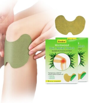
- Mugwort Wormwood Pain Relief Patch for Neck Pain
People Also Ask
- How is sublingual administration different from transdermal? Key Differences & Clinical Uses
- What role does a desiccator play in the moisture content analysis of transdermal patches? Ensure Stability and Safety
- How do transdermal patches improve medication adherence? Enhance Treatment Compliance with Ease
- How does high-purity far-infrared ceramic powder contribute to the efficacy of far-infrared physical therapy patches?
- Can all medications be made into transdermal forms? Understanding the Limits of Skin Delivery