High-precision nanoparticle analyzers are the primary tools for validating the quality and longevity of drug delivery systems like Rutin-loaded transfersomes. By utilizing Dynamic Light Scattering (DLS), these instruments simultaneously monitor the average vesicle size and uniformity (PDI) while measuring Zeta potential to predict and ensure long-term resistance to aggregation.
The Core Stability Principle For flexible transfersomes, stability is not just about chemical composition; it is defined by physical forces. The analyzer verifies that particles possess sufficient electrostatic repulsion—typically a Zeta potential between -17 and -22 mV—to push against one another and prevent the formulation from clumping during storage.
The Critical Role of Physical Characterization
Monitoring Vesicle Size and Distribution
The physical size of a transfersome dictates its effectiveness. To penetrate narrow skin pores, vesicles must be reduced to a nanometer scale, ideally around 170nm.
A high-precision analyzer uses Dynamic Light Scattering (DLS) to confirm that the manufacturing process (often ultrasonic processing) has successfully achieved this target size.
Beyond average size, the analyzer measures the Polydispersity Index (PDI). A low PDI indicates a uniform formulation, which is essential for consistent drug release and skin absorption.
Zeta Potential as a Stability Shield
Zeta potential measures the electrical charge on the surface of the particle. This metric is the most vital indicator of storage stability.
In Rutin-loaded transfersomes, a Zeta potential ranging from -17 to -22 mV provides a barrier of electrostatic repulsion.
Without this repulsion, particles would naturally attract and adhere to one another. The analyzer confirms this charge is present, ensuring the particles remain distinct and suspended rather than aggregating into large, unusable clumps.
Verifying Surface Modifications
Advanced transfersomes often undergo surface modifications, such as the attachment of cell-penetrating peptides, to enhance performance.
The analyzer serves as a validation tool for these modifications. Because these additives alter the surface chemistry, a significant shift in Zeta potential readings acts as a physical indicator that the surface functionalization was successful.
Understanding the Trade-offs
Measurement vs. Manufacture
It is important to distinguish between monitoring stability and creating it. The analyzer diagnoses the state of the formulation, but it cannot correct it.
For example, if the analyzer detects a high PDI (high non-uniformity), it indicates a failure in the upstream processing—such as insufficient cavitation during ultrasonic processing—rather than a flaw in the analyzer itself.
The Limits of Zeta Potential
While a Zeta potential between -17 and -22 mV is ideal for this specific formulation, it is not a universal guarantee of stability for all nanocarriers.
Relying solely on charge without considering particle size (PDI) can lead to false confidence. A formulation may have excellent electrostatic repulsion but still fail if the particle sizes are too irregular to penetrate the skin barrier effectively.
Making the Right Choice for Your Goal
When interpreting data from a nanoparticle analyzer, tailor your focus to your immediate objective:
- If your primary focus is Long-Term Storage: Prioritize Zeta potential readings; ensure they fall strictly within the -17 to -22 mV range to guarantee sufficient repulsion against aggregation.
- If your primary focus is Skin Penetration Efficiency: Prioritize Average Particle Size and PDI; confirm the size is approximately 170nm with a low PDI to ensure the flexibility needed to traverse skin pores.
Accurate analysis transforms raw formulation data into a predictive roadmap for product success.
Summary Table:
| Metric | Target Range | Critical Function for Rutin Transfersomes |
|---|---|---|
| Particle Size | ~170 nm | Ensures effective penetration through narrow skin pores. |
| PDI | Low (Uniform) | Guarantees consistent drug release and absorption. |
| Zeta Potential | -17 to -22 mV | Provides electrostatic repulsion to prevent aggregation. |
| Surface Charge | Variable | Validates successful attachment of cell-penetrating peptides. |
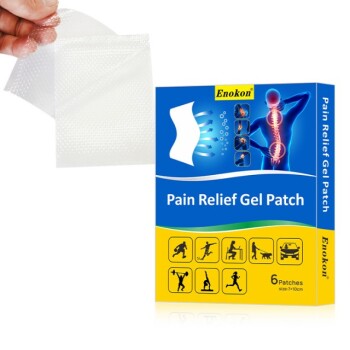
Elevate Your Topical Drug Delivery with Enokon
Precise characterization is the first step toward a successful transdermal product. As a trusted manufacturer and R&D partner, Enokon specializes in high-quality wholesale transdermal patches and custom formulation solutions. From Lidocaine and Menthol pain relief to specialized Eye Protection and Detox patches, we bring pharmaceutical-grade expertise to your brand.
Our value to you:
- Custom R&D: Expert guidance on formulation stability and nanoparticle characterization.
- Scalable Manufacturing: Wholesale production of advanced patches (excluding microneedle technology).
- Proven Efficacy: Products designed for optimal skin penetration and shelf-life stability.
Ready to bring your innovative transdermal solution to market? Contact us today to discuss your custom R&D needs!
References
- Kamlesh Wadher, Milind Umekar. Formulation and Cytotoxic Characterization of Rutin Loaded Flexible Transferosomes For Topical Delivery: Ex-Vivo And In-Vitro Evaluation. DOI: 10.2139/ssrn.4145403
This article is also based on technical information from Enokon Knowledge Base .
Related Products
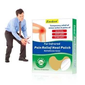
- Mugwort Wormwood Pain Relief Patch for Neck Pain
- Heating Pain Relief Patches for Menstrual Cramps
- Silicone Scar Sheets Patch Transdermal Drug Patch
- Far Infrared Heat Pain Relief Patches Transdermal Patches
- Hydra Gel Health Care Eye Patch
People Also Ask
- What type of pain are pain relief patches best suited for? Find Targeted Relief for Your Aches
- How do pain relief patches compare to oral painkillers? Targeted Relief vs. Systemic Effects
- How should pain relief patches be applied and used? A Guide to Safe & Effective Targeted Relief
- What are pain relief patches and how are they used? A Guide to Safe, Targeted Relief
- Who should consult a healthcare professional before using pain relief patches? Ensure Your Safety with Medical Advice