Quantifying skin irritation and adhesive performance requires a fusion of biochemical staining and high-precision digital analysis. To observe the effects of adhesive tape removal on the stratum corneum, researchers apply a 1% Brilliant Red B solution to dye the skin cells (corneocytes) stripped away by the tape, followed by high-resolution digital microscopic imaging to calculate the exact percentage of the adhesive surface covered by these cells.
This integrated methodology transforms a subjective "skin pull" into objective, calculable data, allowing brands to scientifically validate the gentleness and safety of their adhesive formulations through rigorous R&D.
The Science of Visualization: Staining the Stratum Corneum
Precision Dyeing with Brilliant Red B
The process begins by using a specific staining agent, such as a 1% Brilliant Red B solution, to treat the adhesive tape after it has been removed from the skin. This dye specifically targets the proteins in the captured skin cells, making them highly visible against the adhesive backdrop.
Identifying Cellular Displacement
Without staining, individual skin cells are often translucent and nearly impossible to quantify accurately on an adhesive surface. The staining process provides the necessary contrast, ensuring that every microscopic fragment of the stratum corneum is accounted for during the evaluation phase.
Supporting Custom Formulation R&D
For brand owners, this staining technique is a cornerstone of custom formulation development. It allows our R&D teams to adjust adhesive tackiness and chemical compositions to meet specific "low-trauma" or "sensitive skin" requirements for global markets.
Digital Microscopic Imaging and Data Quantification
High-Resolution Image Capture
Once the cells are stained, a high-resolution digital microscope captures detailed images of the tape’s surface. This technology eliminates human error and provides a permanent, visual record of the product's physical impact on human tissue.
Algorithmic Surface Analysis
Advanced image analysis software then processes these captures to quantify the percentage of cellular coverage. By calculating the ratio of stained cells to the total adhesive area, we generate a "skin stripping index" that serves as a benchmark for product safety.
Ensuring Stringent Quality Control
At an enterprise manufacturing scale, this data-driven approach is vital for Quality Control (QC). It ensures that every high-volume production batch meets the rigorous safety standards required by medical-grade and GMP-certified facilities.
Understanding the Trade-offs and Technical Limitations
Sensitivity to Environmental Variables
While the staining method is highly effective, results can be influenced by skin hydration levels and ambient humidity during the testing phase. Consistent testing environments are mandatory to ensure that the data remains comparable across different product batches.
Resolution vs. Processing Speed
High-resolution microscopic analysis provides deep insight but requires significant processing time and specialized expertise. While this is ideal for turnkey contract R&D, it is often more intensive than the rapid, qualitative "peel tests" used in lower-tier manufacturing.
Limitations of Two-Dimensional Imaging
Digital imaging measures the area of skin removal but does not always account for the depth of the removal. For products targeting deep-tissue adhesion, this method must be supplemented with additional dermatological assessments to provide a 360-degree safety profile.
How to Apply This to Your Project
Aligning Validation with Brand Goals
The level of microscopic validation you require depends heavily on your target demographic and the regulatory requirements of your specific region. Leveraging these scientific insights allows you to market your products with unparalleled transparency and authority.
- If your primary focus is sensitive skin or pediatric care: Prioritize adhesive formulations that demonstrate the lowest percentage of cellular coverage in microscopic staining tests.
- If your primary focus is long-wear industrial or athletic tape: Use this data to find the "sweet spot" where high adhesion is maintained without causing excessive damage to the stratum corneum.
- If your primary focus is global regulatory compliance: Utilize the quantitative data reports from digital imaging as part of your technical dossier to expedite certifications in highly regulated markets.
By integrating microscopic precision with large-scale manufacturing, you ensure your brand delivers products that are as scientifically sound as they are commercially successful.
Summary Table:
| Process Phase | Key Technology | Primary Function | Business Value |
|---|---|---|---|
| Cell Visualization | 1% Brilliant Red B | Dyes proteins in skin cells | High contrast for accurate measurement |
| Data Capture | Digital Microscopy | High-res image acquisition | Eliminates human error & records data |
| Quantification | Analysis Software | Algorithmic surface calculation | Objective safety benchmarks (QC) |
| R&D Application | Custom Formulations | Adjusting adhesive tackiness | Validates "sensitive skin" marketing claims |
Scale Your Brand with Scientifically Validated Adhesives
At Enokon, we combine enterprise-level manufacturing scale with elite R&D prowess to deliver high-performance transdermal solutions. As a trusted manufacturer and OEM/ODM partner, we offer brand owners, distributors, and wholesalers a turnkey path to market-leading products.
Why Partner with Enokon?
- Turnkey R&D: Custom formulations (Lidocaine, Menthol, Capsicum, Herbal, and more) validated by microscopic analysis.
- Massive Capacity: GMP-certified facilities capable of reliable, high-volume delivery for global markets.
- Global Standards: Stringent quality control and comprehensive certifications to ensure regulatory compliance.
- Diverse Portfolio: From pain relief and detox to medical cooling gels (excluding microneedle technology).
Ready to enhance your product lineup with high-margin, low-trauma adhesive technology?
Contact Our Expert Team Today to discuss your custom manufacturing needs!
References
- Michael Turnbull, Amy K. McNulty. Comparison of Medical Tape Performance Using Skin Response Quantitative Measurements on Healthy Volunteers. DOI: 10.7759/cureus.56548
This article is also based on technical information from Enokon Knowledge Base .
Related Products
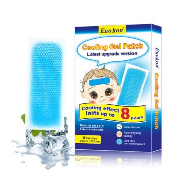
- Medical Cooling Gel Patches for Fever Cooling Patches
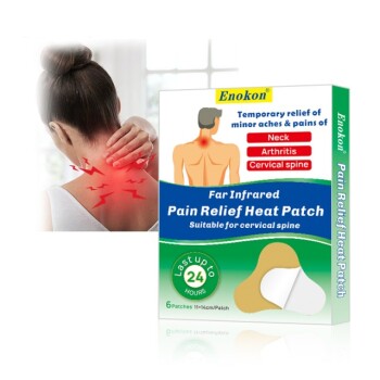
- Far Infrared Deep Heat Relief Patches Medicated Pain Relief Patches
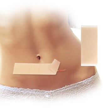
- Silicone Scar Sheets Patch Transdermal Drug Patch
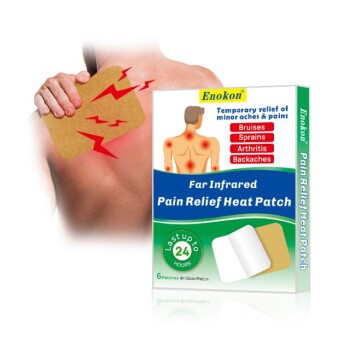
- Far Infrared Heat Pain Relief Patches Transdermal Patches
- Icy Hot Menthol Medicine Pain Relief Patch
People Also Ask
- How does the mechanism of action for medical cooling gel patches facilitate physical temperature reduction? Science Explained
- What role do medical ice packs or cooling gel patches play during the application of high-concentration capsaicin patches?
- What are the advantages of cooling patches over traditional wet towel methods? Modern Fever Relief
- How do cooling patches work to reduce fever? Safe, Drug-Free Relief for All Ages
- What is the physical mechanism of Medical Cooling Gels in relieving skin inflammation? Scientific Insights for OEMs.