High-resolution imaging is the cornerstone of precision R&D for transdermal delivery systems. It allows manufacturers to visualize, quantify, and optimize how patches interact with the skin's microstructure at a cellular level. By providing empirical data on micro-channel formation and drug migration, these technologies ensure that every batch meets the stringent efficacy and safety standards required for global distribution.
High-resolution imaging transforms transdermal patch development from a theoretical exercise into a data-driven manufacturing science. It provides the visual evidence necessary to optimize formulations, validate penetration depth, and ensure the structural integrity of patches at an enterprise scale.
Visualizing Skin Micro-Channel Dynamics
Monitoring Opening and Recovery
High-resolution imaging is used to record morphological changes on the skin surface immediately following patch application. By observing the skin's microscopic morphology after removal, researchers can assess how different concentrations of active components affect the opening and closing of micro-channels. This data is vital for ensuring that skin integrity is maintained while maximizing drug delivery.
Validating Penetration Depth and Migration
Using high-resolution chemical imaging, manufacturers can track the migration paths of chemical enhancers like Lidocaine or Menthol. This technology allows technical teams to see exactly how deep active ingredients penetrate, from the stratum corneum to the deeper dermis. For B2B partners, this provides a visual guarantee that the product will reach the target physiological depth effectively.
Tracking Macromolecule Distribution
Advanced fluorescence microscopy quantifies the distribution of fluorescent markers across skin cross-sections. This allows experts to evaluate the penetration uniformity of macromolecules under various electrical or chemical parameters. Such precision is essential for developing high-end, custom formulations that require exact delivery protocols.
Structural Integrity and Formula Optimization
Nanofiber and Matrix Analysis
Through Scanning Electron Microscopy (SEM), researchers can evaluate the three-dimensional fiber networks of materials like bacterial cellulose. SEM imaging allows for the extraction of quantitative data, such as pore size, fiber diameter, and porosity. These metrics are the core physical parameters used to evaluate drug encapsulation efficiency and diffusion rates in high-performance patches.
Ensuring Uniform Drug Distribution
High-resolution polarizing microscopy is utilized to monitor the distribution of drugs within the polymer matrix. Because specific drugs exhibit birefringence, this equipment detects crystal clusters or aggregates that are invisible to the naked eye. Identifying these irregularities allows for the rapid refinement of drying processes to ensure constant drug release performance across mass-produced batches.
Quantitative Histological Analysis
Beyond surface-level observation, high-definition digital microscopy performs quantitative histological analysis. This includes monitoring fibroblast proliferation and collagen fiber alignment density. These images provide objective evidence of the biological effects on tissue regeneration, verifying the therapeutic potency of the formulation for wound healing or anti-aging applications.
Understanding the Trade-offs
Technical Complexity vs. Production Speed
While high-resolution imaging provides unparalleled data, the process of sample preparation and analysis can be time-intensive. For rapid-turnover OEM projects, balancing the depth of microscopic analysis with aggressive production timelines requires a highly efficient, GMP-certified laboratory workflow. Over-reliance on any single imaging method may lead to a narrow understanding of patch performance.
Data Interpretation and Expert Synthesis
High-resolution images generate massive amounts of raw data that require specialized image processing and expert interpretation. Without a senior technical team to synthesize these findings into actionable manufacturing adjustments, the data remains a luxury rather than a utility. For brand owners, the value lies not just in the images, but in the R&D expertise used to translate those images into a more effective product.
Applying Imaging Data to Your Project Goals
Making the Right Choice for Your Goal
The integration of high-resolution imaging into the manufacturing process ensures that your brand offers a product backed by rigorous scientific validation and consistent quality.
- If your primary focus is Custom Formula Development: Utilize chemical and fluorescence imaging to prove penetration depth and optimize the concentration of active extracts for maximum efficacy.
- If your primary focus is Mass Market Scalability: Rely on SEM and polarizing microscopy to ensure structural uniformity and drug distribution across millions of units, minimizing batch-to-batch variation.
- If your primary focus is Clinical or Therapeutic Claims: Leverage histological imaging to provide objective, visual evidence of skin recovery and tissue regeneration to support your marketing and regulatory filings.
By leveraging these advanced imaging technologies, enterprise-level manufacturers provide the transparency and reliability required to lead in the competitive transdermal market.
Summary Table:
| Imaging Technique | Primary Purpose | Key Manufacturing Benefit |
|---|---|---|
| SEM (Scanning Electron Microscopy) | Analyze fiber networks & porosity | Ensures drug encapsulation efficiency |
| Chemical Imaging | Track migration (e.g., Lidocaine/Menthol) | Validates penetration depth & migration |
| Polarizing Microscopy | Monitor drug distribution in matrix | Prevents crystal clusters for uniform release |
| Fluorescence Microscopy | Quantify macromolecule distribution | Optimizes delivery for custom formulations |
| Digital Histology | Observe tissue regeneration & recovery | Provides empirical data for therapeutic claims |
Partner with Enokon for Scientifically Proven Transdermal Solutions
Elevate your brand with Enokon, a trusted manufacturer and GMP-certified OEM/ODM partner specializing in high-performance transdermal delivery systems. We provide brand owners and wholesalers with massive production capacity and turnkey contract R&D backed by high-resolution imaging data.
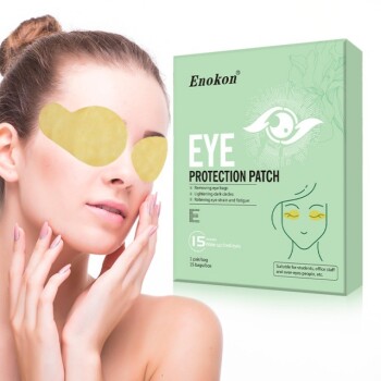
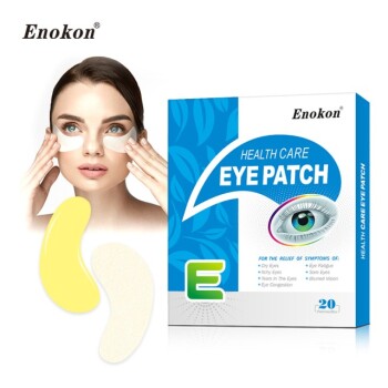
Our comprehensive range of products includes Lidocaine, Menthol, Capsicum, Herbal, and Far Infrared pain relief patches, as well as Eye Protection, Detox, and Medical Cooling Gel solutions (excluding microneedle technology).
Why choose Enokon?
- Custom Formulations: Tailored R&D to meet your specific therapeutic goals.
- Reliable High-Volume Delivery: Massive scale to support global distribution.
- Stringent Quality Control: Advanced microscopic validation for every batch.
Ready to scale your product line with a reliable, high-tech manufacturing partner?
Contact Enokon Today to Get Started
References
- Sachiko Hirobe, Shinsaku Nakagawa. Development and Clinical Study of a Self-Dissolving Microneedle Patch for Transcutaneous Immunization Device. DOI: 10.1007/s11095-013-1092-6
This article is also based on technical information from Enokon Knowledge Base .
Related Products
- Far Infrared Heat Pain Relief Patches Transdermal Patches
- Silicone Scar Sheets Patch Transdermal Drug Patch
- Icy Hot Menthol Medicine Pain Relief Patch
- Menthol Gel Pain Relief Patch
- Mugwort Wormwood Pain Relief Patch for Neck Pain
People Also Ask
- Why is the selection of matrix materials critical when developing customized transdermal patches? Optimize Efficacy
- What are the physical impacts of external heat exposure on the performance and safety of transdermal patches? - Guide
- What role does a skin tolerance scoring system play in the safety evaluation of transdermal patches? Key Safety Metrics
- What are the clinical advantages of local transdermal delivery systems? Unlock Safer, More Effective Pain Relief Solutions
- How does high-purity far-infrared ceramic powder contribute to the efficacy of far-infrared physical therapy patches?