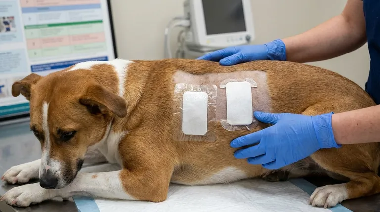
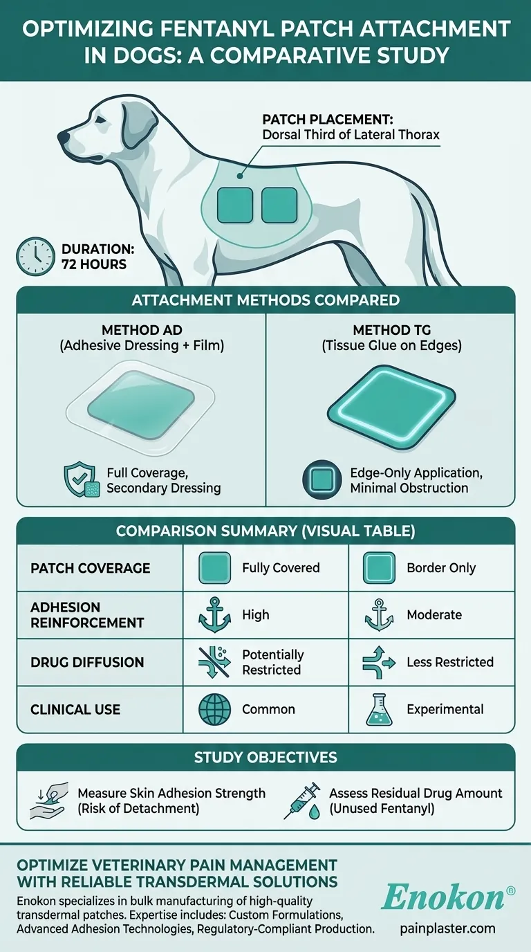
In the study, two matrix-type Fentanyl Patch were applied to each dog for 72 hours to compare attachment methods. Both patches were placed on the dorsal third of the lateral thorax (upper side of the ribcage), with one secured using an adhesive dressing and transparent film (Method AD) and the other with tissue glue applied to the edges (Method TG). The goal was to evaluate how these methods affected patch adhesion and drug retention.
Key Points Explained:
-
Patch Placement Location
- The patches were attached to the dorsal third of the lateral thorax, a stable area with minimal movement, ensuring consistent contact with the skin.
- This location is commonly chosen in veterinary studies due to its accessibility and reduced risk of patch dislodgement from dog activity.
-
Patch Type and Size
- Matrix-type fentanyl patches were used, which rely on drug diffusion through a polymer layer rather than a reservoir system.
- Both patches were equally sized to standardize drug delivery and adhesion comparisons.
-
Attachment Methods Compared
-
Method AD (Adhesive Dressing + Transparent Film):
- A secondary adhesive dressing and transparent film were applied over the patch to reinforce adhesion.
- This mimics common clinical practices where additional securing prevents accidental removal.
-
Method TG (Tissue Adhesive on Patch Edges):
- A tissue adhesive (medical-grade glue) was applied only to the edges of the patch to bond it to the skin.
- This method avoids covering the entire patch, potentially allowing better drug diffusion while maintaining adhesion.
-
Method AD (Adhesive Dressing + Transparent Film):
-
Duration of Application
- Patches remained attached for 72 hours, a standard duration for fentanyl patch studies, ensuring sufficient time to assess adhesion and drug release.
-
Study Objective
- The research aimed to determine whether attachment method influenced:
- Skin adhesion strength (risk of detachment).
- Residual drug amount (how much fentanyl remained unused after 72 hours).
- The research aimed to determine whether attachment method influenced:
This structured approach helps veterinary professionals choose the most effective patch-securing method for pain management in dogs. Would Method TG’s edge-only adhesive compromise adhesion in active animals, or does Method AD’s full coverage unnecessarily restrict drug diffusion? These are practical considerations for clinical use.
Summary Table:
| Aspect | Method AD (Adhesive Dressing + Film) | Method TG (Tissue Glue on Edges) |
|---|---|---|
| Patch Coverage | Full coverage with secondary dressing | Edges only, minimal obstruction |
| Adhesion Reinforcement | High (prevents detachment) | Moderate (relies on edge bonding) |
| Drug Diffusion | Potentially restricted | Less restricted |
| Clinical Use | Common in practice | Experimental, may improve diffusion |
Optimize Veterinary Pain Management with Reliable Transdermal Solutions
At Enokon, we specialize in bulk manufacturing of high-quality transdermal patches, including fentanyl patches for veterinary and human use. Our expertise ensures:
- Custom formulations tailored to your study or clinical needs.
- Advanced adhesion technologies for secure, long-lasting patch performance.
- Regulatory-compliant production for consistent drug delivery.
Need tailored transdermal solutions? Contact our team for R&D support or bulk orders!
Visual Guide
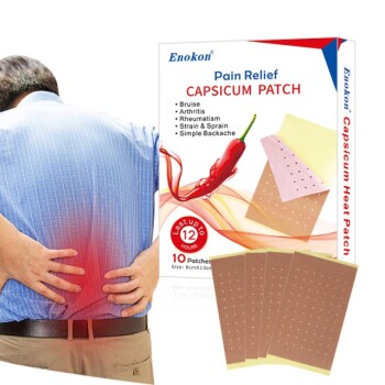
Related Products
- Menthol Gel Pain Relief Patch
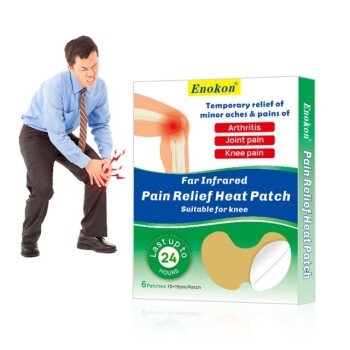
- Far Infrared Heat Pain Relief Patches Transdermal Patches
- Silicone Scar Sheets Patch Transdermal Drug Patch
- Icy Hot Menthol Medicine Pain Relief Patch
- Mugwort Wormwood Pain Relief Patch for Neck Pain
People Also Ask
- Who should consult a healthcare professional before using pain relief patches? Ensure Your Safety with Medical Advice
- What are the potential side effects of pain relief patches? A Guide to Safe Use & Key Risks
- What safety considerations should be kept in mind when using pain relief patches? Ensure Safe & Effective Pain Management
- What are the different types of pain relief patches available? Find the Right Solution for Your Pain
- How do pain relief patches compare to other pain relief methods? Discover Targeted, Long-Lasting Relief