Field Emission Scanning Electron Microscopy (FE-SEM) is the primary tool used to observe the high-resolution microscopic morphology of transdermal drug delivery systems, specifically focusing on surface porosity, interconnected pore structures, and particle dispersion uniformity. It allows R&D teams to verify the structural integrity of polymer matrices—such as PMMA fibers or hydrogels—and ensure that active ingredients are correctly embedded without abnormal aggregation or phase separation.
Core Takeaway: FE-SEM provides the essential microstructural evidence required to validate drug release kinetics and manufacturing consistency, ensuring that transdermal patches meet the stringent quality standards expected in high-volume B2B pharmaceutical production.
Microscopic Precision: Analyzing Porosity and Structure
Surface Porosity and Interconnected Channels
FE-SEM is utilized to characterize the honeycomb-like microporous structures within delivery systems like hydrogels and PMMA fibers. These pores act as the primary channels for solvent penetration and controlled drug release.
By analyzing the pore size distribution, manufacturers can confirm if the matrix meets the specific technical requirements for the target drug's molecular size. This level of detail is critical for ensuring predictable and continuous drug administration.
Variations in Compression Density
Advanced imaging allows for the comparison of compression density between the front and back surfaces of a transdermal patch. These variations can significantly influence how a drug is released once applied to the skin.
This structural data provides a morphological reference used to optimize drug release kinetics. It ensures that the physical properties of the patch support a stable, long-term therapeutic effect.
Verification of Dispersion and Drug Integration
Uniformity of Particle Dispersion
FE-SEM is instrumental in verifying the dispersion uniformity of particles, such as Layered Double Hydroxide (LDH) or Nanostructured Lipid Carriers (NLC), within the polymer matrix. Uniform dispersion is a hallmark of a high-quality, stable formulation.
Visualizing these particles at a nano-scale helps researchers confirm that the spraying or casting processes are performing correctly. This prevents the "clumping" that can lead to inconsistent dosing.
Identification of Drug Crystallization
High-magnification imaging of a patch’s cross-section allows for the visual evaluation of drug crystallization or phase separation. If drug molecules crystallize within the matrix, it can compromise both the efficacy and the mechanical stability of the patch.
Ensuring a smooth, integrated matrix free from macroscopic separation is a key metric for the effectiveness of the physical mixing process. This protects the brand's reputation for product reliability.
Manufacturing Quality and Defect Detection
Detecting Microscopic Structural Flaws
At an enterprise manufacturing scale, FE-SEM serves as a rigorous quality control tool to identify microscopic defects like cracks, bubbles, or abnormal particle aggregation. These flaws are often invisible to the naked eye but can lead to batch failure.
Identifying these issues early in the R&D or pilot phase allows for the optimization of casting processes. This ensures that high-volume production runs maintain a consistent, defect-free quality.
Validating Nano-Scale Loading
For specialized products, such as those involving nano-insulin or silver nanoparticles, FE-SEM confirms that the active ingredients are successfully loaded within the matrix pores. It provides the visual proof of successful "embedding."
This microscopic evidence is essential for GMP-certified facilities to demonstrate that their custom formulations meet the precise design specifications of the client. It builds a foundation of trust between the manufacturer and the brand owner.
Understanding the Trade-offs
High Resolution vs. Sample Alteration
While FE-SEM provides unparalleled resolution, the vacuum environment required for imaging can sometimes alter the state of highly hydrated samples, such as certain hydrogels. This requires specialized preparation techniques to ensure the observed structure reflects the actual product.
Morphological Data vs. Chemical Identity
FE-SEM excels at showing where a particle is and what it looks like, but it does not inherently identify the chemical composition of that particle. Manufacturers often pair FE-SEM with Energy Dispersive X-ray Spectroscopy (EDS) to confirm that the observed particles are indeed the active drug.
Leveraging FE-SEM for Enterprise-Level Product Reliability
How to Apply This to Your Project
Integrating high-resolution microscopic analysis into your product development lifecycle is essential for scaling from a laboratory formula to a mass-marketed transdermal solution.
- If your primary focus is Brand Integrity: Use FE-SEM data as part of your technical dossier to prove the consistency and stability of your custom formulations to regulatory bodies.
- If your primary focus is Manufacturing Efficiency: Utilize cross-sectional SEM imaging to validate that high-speed casting and spraying processes are not introducing microscopic bubbles or structural weaknesses.
- If your primary focus is Innovative R&D: Leverage FE-SEM to explore how modified dextrin or cyclodextrin particles interact at the nano-scale, allowing for the creation of next-generation, controlled-release patches.
By demanding this level of microscopic scrutiny, you ensure that your transdermal products provide the safe, continuous, and reliable delivery that modern healthcare consumers require.
Summary Table:
| Characteristic Observed | FE-SEM Insight Provided | Impact on Product Quality |
|---|---|---|
| Surface Porosity | Pore size & distribution (honeycomb structure) | Ensures controlled drug release kinetics. |
| Particle Dispersion | Visualizing NLC/LDH uniformity at nano-scale | Prevents clumping and ensures consistent dosing. |
| Drug Crystallization | Identification of phase separation/crystals | Guarantees mechanical stability and efficacy. |
| Structural Integrity | Detection of microscopic cracks or bubbles | Minimizes batch failures in high-volume production. |
| Compression Density | Comparison of front vs. back surface density | Optimizes stable, long-term therapeutic delivery. |
Partner with Enokon for Advanced Transdermal Manufacturing
Are you looking to scale your brand with a partner who prioritizes microscopic precision and manufacturing excellence? Enokon is a trusted brand and global manufacturer specializing in wholesale transdermal patches and custom R&D solutions for distributors and B2B resellers.
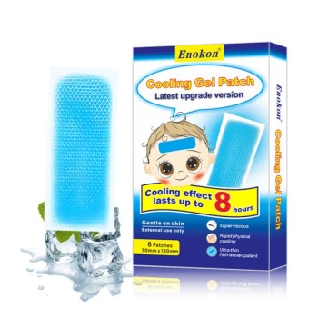
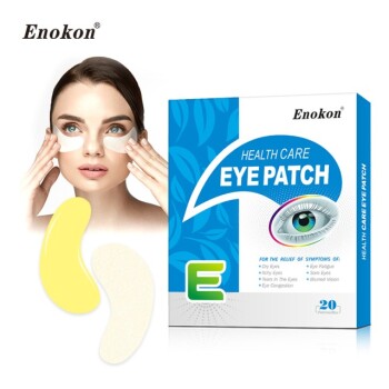
From Lidocaine and Menthol pain relief to Eye Protection, Detox, and Medical Cooling Gel patches, our GMP-certified facilities offer the turnkey contract R&D and massive production capacity your business demands. We leverage advanced analysis to ensure every patch meets stringent quality standards (Note: our range excludes microneedle technology).
Ready to elevate your product line with a reliable OEM/ODM partner?
References
- Thi Thanh Van Tran, Kai Kamada. Enhanced Transdermal Delivery via Electrospun PMMA Fiber Mats Incorporating Ibuprofen-Intercalated Layered Double Hydroxides. DOI: 10.3390/ceramics8040124
This article is also based on technical information from Enokon Knowledge Base .
Related Products
- Far Infrared Heat Pain Relief Patches Transdermal Patches
- Icy Hot Menthol Medicine Pain Relief Patch
- Silicone Scar Sheets Patch Transdermal Drug Patch
- Menthol Gel Pain Relief Patch
- Mugwort Wormwood Pain Relief Patch for Neck Pain
People Also Ask
- Why is the selection of matrix materials critical when developing customized transdermal patches? Optimize Efficacy
- What are the physical impacts of external heat exposure on the performance and safety of transdermal patches? - Guide
- What are the clinical advantages of local transdermal delivery systems? Unlock Safer, More Effective Pain Relief Solutions
- What role does a skin tolerance scoring system play in the safety evaluation of transdermal patches? Key Safety Metrics
- How does high-purity far-infrared ceramic powder contribute to the efficacy of far-infrared physical therapy patches?