The function of a two-compartment diffusion cell in measuring potential gradients is to isolate skin tissue between two independent chambers—simulating the external drug environment and internal physiological conditions—to detect electrochemical changes. By utilizing reference electrodes placed within each chamber, the apparatus measures the transmembrane potential difference generated by the varying diffusion rates of ions. This setup is essential for the quantitative analysis of how ionic drugs navigate the stratum corneum, the skin's outermost barrier.
The core value of this apparatus lies in its ability to translate the physical movement of ions into measurable electrical data. By capturing the potential difference across the skin, researchers can quantify transport mechanisms that standard permeation tests might miss.
The Mechanics of the Setup
Isolating the Environments
The fundamental design of the cell involves sandwiching a biological membrane, typically skin tissue, between two distinct compartments.
One chamber serves as the donor compartment, containing the drug solution or formulation being tested.
The opposing chamber acts as the receptor compartment, containing a physiological saline or buffer solution that mimics the body's internal environment.
The Role of Reference Electrodes
To measure potential gradients specifically, the standard diffusion cell is modified with the installation of reference electrodes in both the donor and receptor chambers.
These electrodes are the critical sensors that detect electrical variances between the two isolated environments.
They provide a continuous readout of the voltage difference across the skin barrier, which serves as a proxy for ion movement.
Simulating Physiological Conditions
While the electrodes measure electricity, the physical apparatus must maintain biological realism to ensure accurate data.
As noted in standard diffusion cell protocols, the system typically employs a circulating water bath to maintain the skin surface temperature (often between 32.5°C and 37°C).
Additionally, magnetic or electromagnetic stirring is used in the receptor chamber to ensure the medium remains uniform, preventing stagnant layers that could skew diffusion rates.
Understanding Potential Gradients
Ion Diffusion and Voltage Generation
The potential gradient is not an inherent property of the cell but a result of ion diffusion kinetics.
As ionic drug molecules penetrate the skin, they move at different rates compared to the counter-ions in the system.
This disparity in movement speed creates a charge separation across the membrane, generating the transmembrane potential difference detected by the electrodes.
Analyzing the Stratum Corneum
The primary barrier to transdermal delivery is the stratum corneum, the dense outer layer of the skin.
This apparatus allows researchers to focus specifically on how this layer interacts with charged molecules.
By correlating the measured potential difference with the known concentration of the drug, researchers can mathematically model the mobility and permeability of the drug within this specific tissue layer.
Operational Considerations and Trade-offs
Sensitivity to Experimental Conditions
The measurement of potential gradients is highly sensitive to external factors.
Slight variations in temperature or pH levels in the receptor fluid can alter ionization states, potentially creating noise in the electrical data.
Precise calibration of the reference electrodes is non-negotiable; establishing a stable baseline before introducing the drug is critical for validity.
In Vitro Limitations
While this method provides excellent quantitative data on ionic movement, it remains an in vitro simulation.
It creates a controlled environment that mimics the steady-state flux of drugs entering the circulatory system, but it cannot perfectly replicate the dynamic blood flow and clearance mechanisms of a living organism.
Researchers must account for these differences when extrapolating lab results to potential clinical outcomes.
Applying This Methodology to Your Research
If you are designing a transdermal study, choose your apparatus based on the specific mechanism of transport you need to evaluate.
- If your primary focus is analyzing ionic mobility and electrical interactions: Select a two-compartment cell equipped with reference electrodes to capture transmembrane potential differences.
- If your primary focus is general drug accumulation and lag time: Utilize a standard vertical Franz diffusion cell to measure the cumulative concentration of the drug in the receptor fluid over time.
By aligning the apparatus with your specific analytical goals, you ensure that the data collected accurately reflects the physical and chemical realities of your drug delivery system.
Summary Table:
| Feature | Function in Two-Compartment Cell |
|---|---|
| Donor Compartment | Holds the drug formulation/solution being tested |
| Receptor Compartment | Mimics physiological environment with buffer solution |
| Reference Electrodes | Detect electrical variances and transmembrane potential |
| Stratum Corneum | Acts as the membrane barrier for ion diffusion analysis |
| Stirring & Temperature | Ensures medium uniformity and biological realism (32.5-37°C) |
Elevate Your Transdermal R&D with Enokon
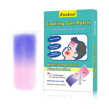
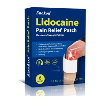
Are you looking to optimize your drug delivery formulations? As a trusted manufacturer and R&D specialist, Enokon provides comprehensive wholesale solutions for transdermal patches. We specialize in high-quality products including Lidocaine, Menthol, Capsicum, Herbal, and Far Infrared pain relief patches, alongside Eye Protection, Detox, and Medical Cooling Gel patches (excluding microneedle technology).
Partner with us for custom R&D and reliable manufacturing that brings your product to market efficiently. Contact us today to discuss your project!
References
- Fumihiko Ikemoto, Kiyoshi Kanamura. Transdermal drug delivery by using electronic potential for driving force. DOI: 10.1254/fpj.137.182
This article is also based on technical information from Enokon Knowledge Base .
Related Products
- Mugwort Wormwood Pain Relief Patch for Neck Pain
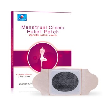
- Heating Pain Relief Patches for Menstrual Cramps
- Silicone Scar Sheets Patch Transdermal Drug Patch
- Far Infrared Heat Pain Relief Patches Transdermal Patches
- Cooling Fever Patches Color Change Cold Fever Patch
People Also Ask
- What are pain relief patches? Discover Targeted, Drug-Free Pain Management Solutions
- What are pain relief patches and how are they used? A Guide to Safe, Targeted Relief
- How do pain relief patches compare to oral painkillers? Targeted Relief vs. Systemic Effects
- What safety considerations should be kept in mind when using pain relief patches? Ensure Safe & Effective Pain Management
- How should pain relief patches be applied and used? A Guide to Safe & Effective Targeted Relief