Hematoxylin and Eosin (H&E) staining is the indispensable "gold standard" for evaluating the skin safety and histopathological impact of transdermal medicinal products. This laboratory technique provides a high-resolution microscopic view of skin tissue to verify that delivery enhancers or delivery devices do not damage the biological skin barrier. By staining cell nuclei and cytoplasm, researchers can identify inflammatory infiltration, tissue necrosis, or structural degradation, ensuring the formulation meets stringent global safety standards before high-volume production.
H&E staining serves as the definitive diagnostic tool for verifying skin integrity and identifying potential toxicities in transdermal drug development. For enterprise-level manufacturers and brand owners, it provides the essential morphological evidence required to bridge the gap between innovative drug delivery and regulatory compliance.
Validating Skin Barrier Integrity and Safety
Microscopic Structural Analysis
H&E staining allows for precise observation of the epidermal structure to ensure it remains intact after the application of gels, patches, or high-voltage pulses. This is vital for confirming that penetration enhancers are facilitating drug delivery without permanently compromising the skin's natural defense.
Identifying Cellular-Level Damage
The process reveals critical indicators of damage, such as epidermal vacuolar degeneration or severe exfoliation. These insights allow R&D teams to refine custom formulations at the laboratory stage before they move into massive production cycles.
Monitoring Dermal Congestion
By staining skin sections from the application site, technicians can check for inflammatory cell infiltration or congestion in the dermis. This ensures that the transdermal product is non-irritating and safe for long-term consumer use.
R&D Optimization and Mechanism of Action
Mapping Penetration Depth
Beyond safety, H&E staining provides the visual contrast needed to see the distribution path of drugs in subcutaneous tissues. This data is essential for optimizing parameters like injection pressures or laser energy in advanced, turnkey transdermal systems.
Confirming Absorption Mechanisms
High-magnification observation reveals how enhancers alter the stratum corneum and increase intercellular spaces. This evidence provides a scientific foundation for the product's efficacy, which is a significant value-add for B2B resellers and brand owners.
Visualizing Micro-Ablation
In products involving physical enhancement (like microneedles or lasers), H&E staining clearly reveals the depth of ablation craters. This allows for the visual and quantitative observation of tissue removal, ensuring the delivery mechanism is both effective and controlled.
Risk Mitigation in Safety Assessment
Differentiating Chemical vs. Immune Reactions
H&E staining helps determine if a skin reaction is caused by direct chemical toxicity or an inflammation-mediated immune response. Distinguishing between these two is critical for navigating the clinical trial process and managing long-term brand liability.
Detecting Sub-clinical Inflammation
Even when skin appears normal to the naked eye, H&E can detect perivascular lymphocyte infiltration in the dermis. This ensures that "invisible" irritants are identified early, protecting the reputation of the final brand in the global market.
Establishing Histological Evidence
For brands seeking global certifications, H&E provides the histological evidence required by regulatory bodies. It confirms that the transdermal enhancement process complies with international clinical safety standards.
Understanding the Trade-offs
The Processing Lead Time
While H&E is the gold standard, the process requires formaldehyde fixation, paraffin embedding, and microtomy. This multi-step sequence requires a GMP-certified facility with significant throughput capacity to prevent bottlenecks in the R&D timeline.
Destructive Testing Limitations
H&E staining is a destructive testing method, meaning the specific tissue sample used for the stain cannot be reused for other tests. Large-scale manufacturers must account for this by maintaining high-volume sample protocols during the R&D phase.
Subjective Interpretation Risks
Histopathological analysis relies on expert observation of morphological changes. Partnering with a manufacturer that possesses a sophisticated R&D infrastructure ensures that microscopic findings are interpreted accurately and consistently.
How to Apply This to Your Project
When evaluating a manufacturing partner for transdermal products, prioritize their ability to provide comprehensive histopathological data. This ensures your brand is backed by scientific rigour and regulatory-ready safety profiles.
- If your primary focus is Regulatory Compliance: Ensure your OEM partner uses H&E staining as a standard protocol to provide the histological evidence needed for GMP and global certifications.
- If your primary focus is Product Efficacy: Use H&E data to verify the penetration depth and absorption mechanisms of your custom formulations to justify premium market positioning.
- If your primary focus is Risk Management: Demand H&E analysis to rule out sub-clinical inflammation or long-term dermal damage, protecting your brand from future liability.
By integrating rigorous H&E staining into the development cycle, brand owners can confidently scale innovative transdermal solutions that are both highly effective and demonstrably safe.
Summary Table:
| Key Role of H&E Staining | Benefit for Brand Owners & Resellers | Technical Insight Provided |
|---|---|---|
| Skin Integrity Verification | Minimizes liability by proving the skin barrier remains intact. | Visualizes epidermal structure and cell layers. |
| Inflammation Detection | Identifies sub-clinical irritation before market launch. | Detects perivascular lymphocyte infiltration. |
| Regulatory Evidence | Provides necessary histological proof for GMP/global certifications. | Official diagnostic documentation for safety files. |
| R&D Optimization | Justifies premium pricing through proven absorption mechanisms. | Maps penetration depth in subcutaneous tissues. |
| Risk Mitigation | Distinguishes between chemical toxicity and immune responses. | Analysis of tissue necrosis and structural damage. |
Bring Your Transdermal Innovation to Market with Confidence
Partner with Enokon, your trusted OEM/ODM manufacturer for high-volume, medical-grade transdermal solutions. We combine massive production capacity with stringent R&D protocols, including H&E staining, to ensure your custom formulations are both effective and safety-verified for global markets.
Why Choose Enokon?
- Turnkey R&D: Custom formulations and histological evidence to support your brand's claims.
- Global Compliance: GMP-certified facilities ensuring your products meet international regulatory standards.
- Reliable High-Volume Supply: Massive scale to support distributors and B2B resellers worldwide.
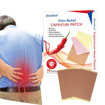
- Diverse Product Range: Specialized in pain relief (Lidocaine, Menthol, Capsicum), Herbal, Eye Protection, and Medical Cooling patches (excluding microneedle technology).
Ready to scale your product line with a partner that prioritizes scientific rigor and manufacturing excellence?
Contact Our Expert Team Today
References
- Heba F. Salem, Hanan O. Farouk. Novel Enhanced Therapeutic Efficacy of Dapoxetine HCl by Nano-Vesicle Transdermal Gel for Treatment of Carrageenan-Induced Rat Paw Edema. DOI: 10.1208/s12249-020-01656-6
This article is also based on technical information from Enokon Knowledge Base .
Related Products
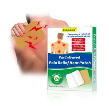
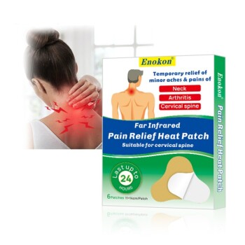
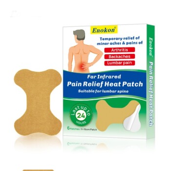
- Far Infrared Heat Pain Relief Patches Transdermal Patches
- Silicone Scar Sheets Patch Transdermal Drug Patch
- Icy Hot Menthol Medicine Pain Relief Patch
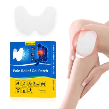
- Menthol Gel Pain Relief Patch
- Mugwort Wormwood Pain Relief Patch for Neck Pain
People Also Ask
- Why is the selection of matrix materials critical when developing customized transdermal patches? Optimize Efficacy
- What role does a skin tolerance scoring system play in the safety evaluation of transdermal patches? Key Safety Metrics
- What are the clinical advantages of local transdermal delivery systems? Unlock Safer, More Effective Pain Relief Solutions
- What are the physical impacts of external heat exposure on the performance and safety of transdermal patches? - Guide
- How does high-purity far-infrared ceramic powder contribute to the efficacy of far-infrared physical therapy patches?