Scanning Electron Microscopy (SEM) serves as the primary validation tool for visualizing the internal architecture of composite hydrogel patches. It allows for the direct observation of internal pore distribution and network crosslinking density within freeze-dried hydrogels. Specifically, SEM confirms whether nanoparticles are uniformly embedded in the matrix and verifies if distinct polymer components—such as agar, gelatin, and sodium polyacrylate—have successfully formed a cohesive, interpenetrating network.
Core Insight: SEM transforms theoretical formulation data into physical evidence. It proves whether your manufacturing process successfully created the specific microscopic network required to control drug release and maintain structural integrity.
Visualizing Internal Architecture
Verifying Network Crosslinking
The primary function of SEM in this context is to reveal the crosslinking density of the hydrogel. By imaging the freeze-dried structure, you can assess how tightly the polymer chains are interconnected.
A dense, uniform network typically indicates a robust formulation capable of sustained drug delivery. Conversely, irregular or loose networks may suggest inconsistencies in the polymerization process.
Confirming Composite Integration
Composite hydrogels often rely on blending different polymers, such as agar and gelatin, to achieve specific mechanical properties.
SEM imaging allows you to see if these components have formed a true interpenetrating network structure. This confirms that the materials have physically or chemically integrated rather than remaining as separate, phase-separated regions which could weaken the patch.
Assessing Payload Distribution
Nanoparticle Embedding
For patches utilizing advanced delivery systems, SEM is critical for verifying nanoparticle uniformity.
You can directly observe whether nanoparticles are evenly distributed throughout the polymer matrix or if they have agglomerated in specific areas. Uniform embedding is essential for ensuring consistent therapeutic dosage across the entire patch.
Detecting Drug Crystallization
Beyond nanoparticles, SEM helps distinguish the physical state of the drug within the patch.
High-resolution imaging can reveal if the drug exists as a molecular dispersion (ideal for some formulations) or if there is crystalline precipitation. Detecting unwanted crystals is vital, as they can significantly alter dissolution rates and bioavailability.
Connecting Microstructure to Performance
Predicting Drug Release Behavior
The micro-morphology revealed by SEM directly correlates to how a drug will exit the patch.
By analyzing the pore size and connectivity, researchers can predict diffusion pathways. For example, a sponge-like or honeycomb structure observed after release experiments confirms that the drug diffused through a swollen matrix, helping to characterize the release mechanism (e.g., linear release vs. burst release).
Estimating Permeability and Absorption
SEM allows for the measurement of specific pore dimensions, often typically ranging from 45 to 120 µm in these applications.
These structural details are essential for determining the patch's moisture absorption performance and breathability. The presence of uniform micropores suggests the patch can effectively absorb biological fluids while maintaining skin compatibility.
Understanding the Trade-offs
The "Freeze-Dried" Artifact
It is critical to remember that SEM typically images hydrogels in a freeze-dried state. While this preserves the 3D network structure for observation, it is a static representation of the material in a dry environment, not the hydrated state it will assume on the skin.
Manufacturing Defects
While SEM confirms successful structures, it is equally valuable for spotting failures. It serves as a quality control checkpoint to detect micro-cracks, surface irregularities, or uneven film thickness. These defects are early indicators of potential mechanical failure or inconsistent drug delivery in the final product.
Making the Right Choice for Your Goal
To maximize the value of SEM data for your specific development phase, focus on these indicators:
- If your primary focus is Controlled Release: precise measurement of pore size and network density is required to predict diffusion rates and prevent dose dumping.
- If your primary focus is Formulation Stability: Look for the uniform embedding of nanoparticles and the absence of phase separation between polymer components to ensure shelf-life integrity.
- If your primary focus is Manufacturing Quality: Use SEM to scan for surface micro-cracks and crystal precipitation, which indicate processing errors or saturation limits.
Ultimately, SEM provides the visual proof that your chemical formulation has successfully translated into a functional physical structure.
Summary Table:
| SEM Analysis Feature | Key Information Provided | Impact on Patch Performance |
|---|---|---|
| Network Crosslinking | Density and uniformity of polymer chains | Structural integrity and sustained drug release |
| Nanoparticle Embedding | Distribution uniformity vs. agglomeration | Consistent therapeutic dosage across the patch |
| Pore Morphology | Pore size (e.g., 45-120 µm) and connectivity | Breathability, moisture absorption, and diffusion rates |
| Component Integration | Presence of interpenetrating networks | Mechanical strength and formulation stability |
| Defect Detection | Micro-cracks and drug crystallization | Quality control and prevention of mechanical failure |
Elevate Your Product Quality with Enokon’s Manufacturing Expertise
At Enokon, we bridge the gap between advanced microstructure analysis and large-scale production. As a trusted manufacturer specializing in wholesale transdermal patches and custom R&D solutions, we ensure that every formulation—from Lidocaine and Menthol pain relief to specialized Herbal and Detox patches—meets the highest standards of structural integrity.
Our expertise in polymer matrices (excluding microneedle technology) allows us to deliver high-performance products including:
- Medical Cooling Gel & Eye Protection Patches
- Advanced Pain Relief (Capsicum, Far Infrared)
- Custom R&D for Interpenetrating Polymer Networks
Partner with a leader in transdermal drug delivery to ensure your products offer superior absorption and stability. Contact us today to discuss your custom R&D needs!
References
- Shasha Wang, Jianping Liu. Enhanced intradermal delivery of Dragon's blood in biocompatible nanosuspensions hydrogel patch for skin photoprotective effect. DOI: 10.1111/jocd.15515
This article is also based on technical information from Enokon Knowledge Base .
Related Products
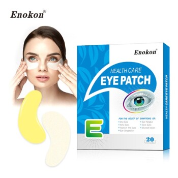
- Hydra Gel Health Care Eye Patch
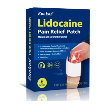
- Lidocaine Hydrogel Pain Relief Patch for Pain Relief
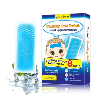
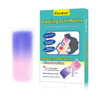
- Medical Cooling Gel Patches for Fever Cooling Patches
- Menthol Gel Pain Relief Patch
- Mugwort Wormwood Pain Relief Patch for Neck Pain
People Also Ask
- What are hydrogel patches made of? Key Ingredients & Benefits Explained
- When should a doctor be consulted regarding the use of this patch? Key Safety Guidelines
- How often can hydrogel eye patches be used? Optimize Your Under-Eye Care Routine
- How should hydrogel eye patches be applied? Maximize Absorption for Brighter, Firmer Skin
- What are some active ingredients in hydrogel eye patches and their purposes? Key Benefits Explained