Histopathological examination using optical microscopy is the definitive diagnostic tool for verifying both the clinical safety and manufacturing precision of transdermal delivery systems. It enables researchers to identify microscopic anatomical changes in skin tissue—such as cell necrosis, edema, or barrier impairment—ensuring that the delivery process is effective without causing irreversible damage or irritation.
Core Takeaway: For brand owners and B2B partners, histopathology serves as the ultimate quality-control benchmark, providing visual and quantitative proof that a transdermal formulation is safe for consumers and manufactured with high uniformity.
Verifying Clinical Safety and Skin Integrity
Identifying Cellular-Level Irritation
Optical microscopy, often utilizing H&E (Hematoxylin and Eosin) staining, is the gold standard for assessing how a formula affects skin layers.
Technical experts use these examinations to detect basal layer edema, epidermal thinning, or the disruption of dermal collagen fibers.
By identifying these pathological changes, manufacturers can establish clinical safety thresholds and ensure that any induced skin changes are reversible.
Distinguishing Drug Effects from Tissue Damage
A primary purpose of this examination is to determine if a drug’s performance is due to its inherent characteristics or a change in the skin’s state.
Researchers can perform semi-quantitative assessments on skin tissue to see if the transdermal process itself has compromised the skin barrier.
This distinction is critical for R&D validation, allowing brand owners to claim their products are non-irritating and dermatologically sound.
Validating Manufacturing Precision and Uniformity
Monitoring Patch Matrix Reliability
Beyond tissue analysis, optical microscopy is used for the macro and micro inspection of finished transdermal patch laminates.
High-resolution observation allows technicians to identify undissolved penetration enhancer crystals, impurities, or unevenly distributed drug areas.
This process ensures the physical uniformity of the patch matrix, which is vital for maintaining consistent dosage across high-volume production runs.
Evaluating Drug Particle Morphology
Using transmission optical microscopy with backlighting, manufacturers can observe the spatial distribution of drug particles within a polymer matrix.
This helps identify drug recrystallization or abnormal aggregation that may have occurred during the manufacturing process.
Verifying these distribution states validates that the production technique has achieved the homogeneity required for GMP-certified, enterprise-level distribution.
Quantitative R&D for Enhanced Efficacy
Measuring Therapeutic Markers
Professional biological microscopes with 400x magnification allow for the quantitative evaluation of skin healing and regeneration.
Technicians measure epidermal thickness, count inflammatory cells, and observe the expression of specific proteins like COX-2.
These objective criteria provide B2B clients with data-backed evidence that their custom formulations effectively promote dermal remodeling and scarless healing.
Optimization of Emulsion Systems
In transdermal emulsions, microscopy is used to observe droplet morphology and distribution uniformity.
By comparing images of emulsions formed under different processing parameters, R&D teams can refine shear speeds to achieve ideal droplet size.
This refinement ensures a stable delivery system that maintains its integrity from the factory floor to the end consumer.
Understanding the Trade-offs
Destructive Testing Requirements
Histopathological examination is a destructive process that requires fixing, sectioning, and staining skin samples.
While it provides the most detailed visual evidence, it cannot be performed in real-time on live human subjects during the manufacturing flow.
Expert Interpretation Necessity
The value of optical microscopy is heavily dependent on the skill of the pathologist or technical researcher.
Automated image analysis can assist, but identifying subtle tissue degradation or spongiosis requires highly trained eyes to avoid misinterpretation of artifacts.
How to Apply This to Your Project
Making the Right Choice for Your Goal
- If your primary focus is brand safety and risk mitigation: Request comprehensive H&E staining reports to prove your formulation does not cause irreversible skin barrier impairment.
- If your primary focus is product shelf-life and stability: Utilize optical microscopy to inspect for recrystallization or enhancer precipitation in the adhesive matrix over time.
- If your primary focus is clinical superiority: Leverage quantitative microscopic indicators, such as epidermal thickness and fibroblast counts, to differentiate your product in a competitive market.
Incorporating rigorous histopathological standards ensures that your transdermal products meet the highest benchmarks for safety, efficacy, and manufacturing excellence.
Summary Table:
| Application Category | Primary Purpose | Key Microscopic Metrics |
|---|---|---|
| Clinical Safety | Detect irritation & cell necrosis | H&E staining, basal layer edema |
| Manufacturing Quality | Ensure patch matrix uniformity | Drug recrystallization, particle distribution |
| R&D Validation | Quantify therapeutic efficacy | Epidermal thickness, inflammatory cell count |
| Formulation Stability | Optimize emulsion systems | Droplet morphology, shear speed results |
Partner with Enokon for High-Precision Transdermal Solutions
Are you looking for a manufacturing partner that prioritizes scientific rigor and GMP-certified quality? Enokon is a trusted brand and manufacturer specializing in high-volume wholesale and custom R&D for the global market. We help brand owners, distributors, and B2B resellers bring safe, effective products to market with data-backed reliability.
Our Core Capabilities & Product Range:
- Turnkey R&D: Custom formulations and semi-quantitative validation to ensure non-irritating, high-performance delivery.
- Massive Capacity: Stringent quality control across high-volume production runs for consistent dosage.
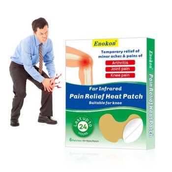
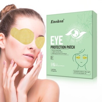
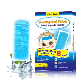
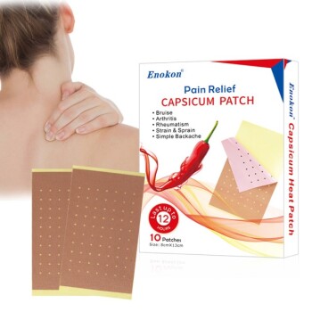
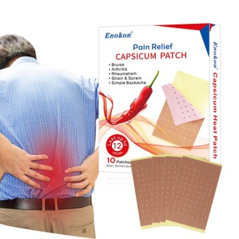
- Diverse Portfolio: Expert manufacturing of Lidocaine, Menthol, Capsicum, Herbal, and Far Infrared pain relief patches, plus Eye Protection and Medical Cooling Gel patches (excluding microneedle technology).
- Global Standards: GMP-certified facilities ensuring your products meet international regulatory benchmarks.
Ready to elevate your product line with enterprise-level manufacturing?
Contact Enokon Today for Custom R&D and Wholesale Quotes
References
- Johannes P Venter, Colleen Goosen. A comparative study of an in situ adapted diffusion cell and an in vitro Franz diffusion cell method for transdermal absorption of doxylamine. DOI: 10.1016/s0928-0987(01)00110-5
This article is also based on technical information from Enokon Knowledge Base .
Related Products
- Far Infrared Heat Pain Relief Patches Transdermal Patches
- Silicone Scar Sheets Patch Transdermal Drug Patch
- Icy Hot Menthol Medicine Pain Relief Patch
- Menthol Gel Pain Relief Patch
- Mugwort Wormwood Pain Relief Patch for Neck Pain
People Also Ask
- What role do transdermal patches play in improving skin lesions? Discover How Stabilization Prevents Pressure Sores
- What is the purpose of vacuum filtration for polymer solutions? Ensuring Quality in Transdermal Patch Manufacturing
- What are the disadvantages of transdermal drug delivery? Key Limitations and Patient Challenges
- How do transdermal patches improve medication adherence? Enhance Treatment Compliance with Ease
- How is sublingual administration different from transdermal? Key Differences & Clinical Uses